PRISM

A Panel-based Spatial Transriptomic Imaging Method for ALL Biologists

PRISM Code

Main Content

BARCODE EXPANSION

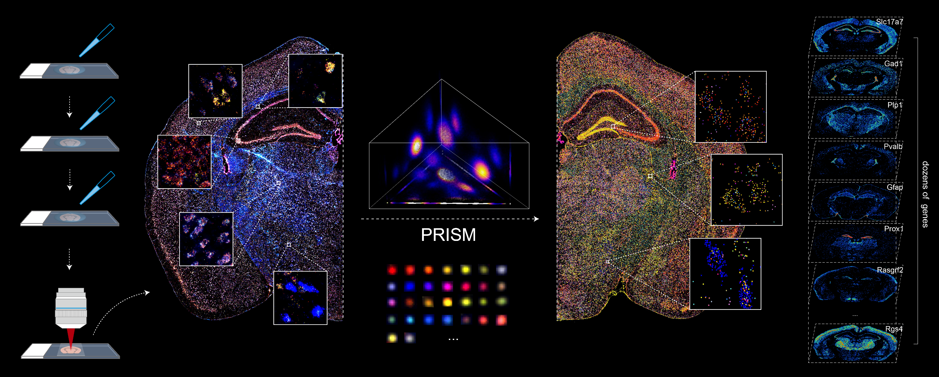
Barcoded padlock probes are designed to hybridize targeted gene transcripts and are amplified locally into clones via rolling circle amplification. Each barcode contains segments corresponding to a spectral channel, taking the spectral combination (both wavelength and intensity) for coding.

The intensity information was encoded in the ratio between fluorescently labeled and unlabeled secondary probes. Using four channels, we have experimentally verified the barcode multiplexity of up to 64 genes in a single imaging round.

CODING PRINCIPLE

Variations in the sizes of amplified nanoballs could make specific codes indistinguishable in spectral color space. To better understand the discriminability of barcodes, we introduced the concept of ‘radius vector’ in color space, and only angularly distinct barcode vectors were considered legitimate.

To ensure ample dispersion of barcode vectors, we chose the plane (Ch1+Ch2+Ch3 = 4) in the coding space as the constraint, reaching an optimal differentiability of intersected barcodes by calculating the angles between any two barcode vectors and eliminating the crosstalk between barcodes.
PRISM Versatility

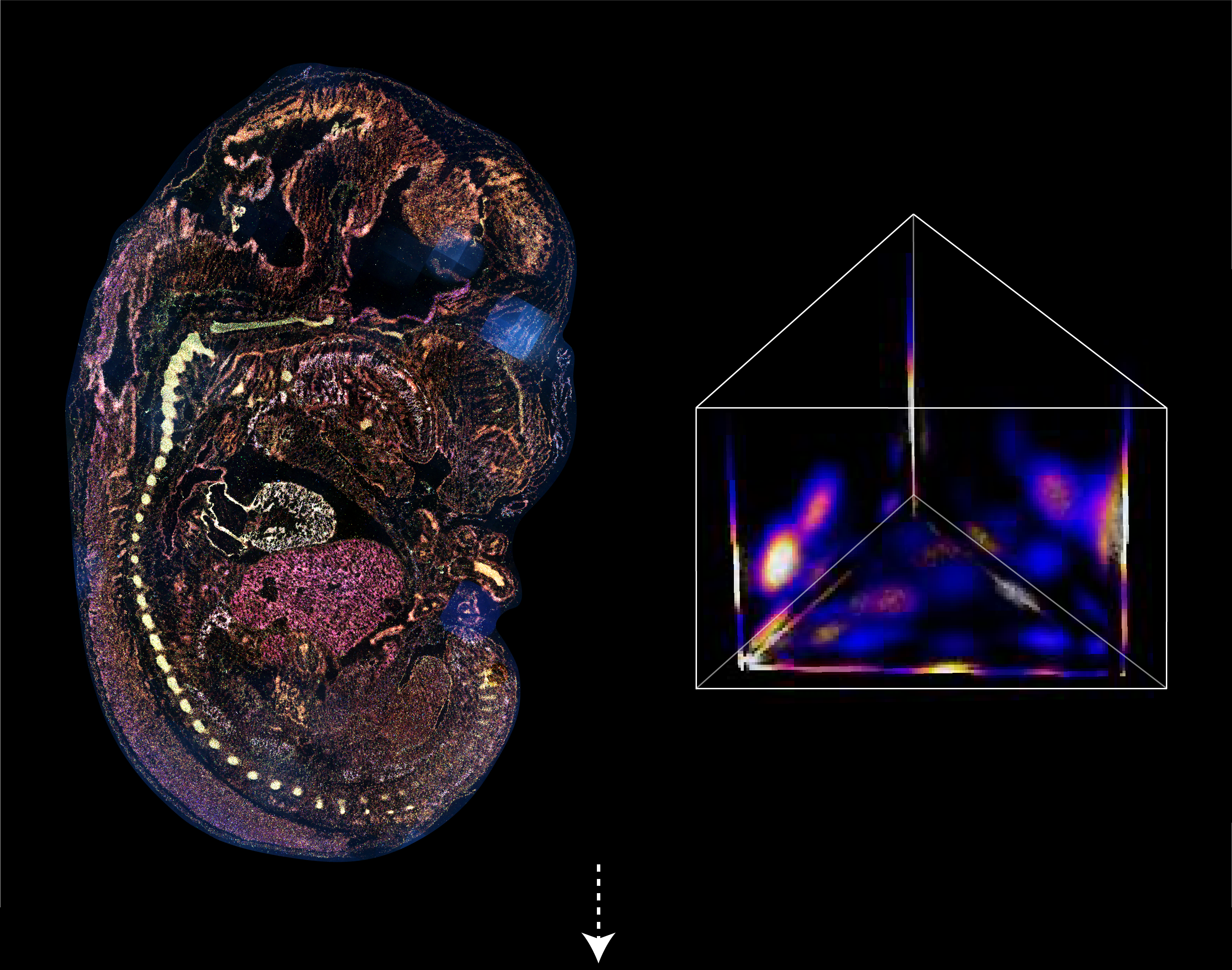
Mouse Embryo

A panel of 30 marker genes was designed to target at various tissues and organs during embryonic development (E13.5 mouse).

Human Hepatocellular Carcinoma

A hepatitis B virus (HBV) positive hepatocellular carcinoma sample was profiled. The panel includes 31 selected markers associated with cell proliferation, liver tumor cells, cancer-associated fibroblasts, endothelial cells, and immune cells, in addition to a probe targeting a 28-bp sequence encoding the HBV core protein.

PRISM 3D Application

3D in-situ GENE IMAGING

PRISM can be applied to intact 3D cell composition in thick tissues. Here are the results from a 100-µm thick mouse brain slice, preserving the 3D structure with high precision and accuracy. Single-round imaging avoids repetitive macromolecule penetration within thick tissues and effectively circumvents the challenges of fluorescent spot alignment and registration across multiple imaging sessions.

The detailed morphology and distribution of marker genes within respective 3D areas offer a new dimension than conventional observations in 2D experiments, bringing new insights into biology and pathological applications.

PRISM 64-plex panel

Col2a1

Acta2

Neurod1

Cldn7

Emx1

Cdx2

Akr1b7

Gad2

Hox5a

Ptix2

Nkx2-1

Hoxb8

Smpx

Afp

Dlx1

En2

Pln

Spp1

Myl7

Tagln

Myl1

Apoa1

Tbx5

Krt5

Robo2

Runx2

Stmn2

Tnnt2

Mef2c

Wnt5a

Foxc2

Ptix1

Ptgds

Hoxd11

Sftpc

Dmrt2

Hba-X

Col11a1

Fabp7

Ttr

Sncg

Alx1

Shox2

Otx2

Sox2

Ttn

Gjb2

Dach1

Shisa3

Myh3

Myh11

Cnpy1

Gabrp

Nppc

Pax6

Actc1

Cdh1

Bmp4

Wwp2

Nkx2-5

Htr2c

Myh6

Myl2

Hand2

Code and Data

Preprint will be available on bioRxiv soon

For more detail information please visit raw data and source code of PRISM in github.

Contact us please email to yanyi@pku.edu.cn

Views: 14164